tibiofemoral joint anatomy
anatomy of Knee joint(膝関節の解剖)(関節、靱帯) | トレンドの樹. 9 Pictures about anatomy of Knee joint(膝関節の解剖)(関節、靱帯) | トレンドの樹 : Knee joint: anatomy, ligaments and movements | Kenhub, Flashcards Table on Anatomy and Physiology Lab 2 and also Normal Knee X-rays | Bone and Spine.
Anatomy Of Knee Joint(膝関節の解剖)(関節、靱帯) | トレンドの樹
 trendnoki.com
Image Result For Gastrocnemius Origin And Insertion | Muscle Anatomy
 www.pinterest.com
gastrocnemius joint insertion origin muscle knee anatomy foot tibiofemoral anterior calf origins serratus body ligaments tibia nerve chandie christina ed
Knee Radiograph (an Approach) | Radiology Reference Article
 radiopaedia.org
knee radiopaedia ray rays radiograph effusion interpretation approach fat suprapatellar lateral pads annotated radiology anterior figure case jeremy jones dr
Normal Knee X-rays | Bone And Spine
 boneandspine.com
knee lateral normal medial ray rotated rays bone xray xr superimpose anatomy condyles evaluation adult differentiating normally slightly absolutely between
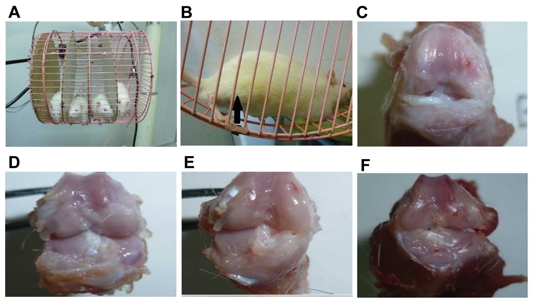
Protective Effect Of Lentivirus-mediated SiRNA Targeting ADAMTS-5 On
 www.spandidos-publications.com
rat osteoarthritis joint cartilage degradation cage macroscopic ijmm targeting sirna lentivirus mediated adamts protective effect analysis mobilization spandidos publications
Презентация на тему: "Knee Dislocation KazNMU 2 топ. Anatomy
 www.myshared.ru
Knee Joint Cartilage Segments Assessed In This Study. LFC, Lateral
 www.researchgate.net
cartilage lfc ltp condyle mfc segments assessed femoral mtp medial tibial plateau
Flashcards Table On Anatomy And Physiology Lab 2
 www.proprofs.com
tibiofemoral joint anatomy flashcards medial knee physiology lab tibial ligament table transverse lateral
Knee Joint: Anatomy, Ligaments And Movements | Kenhub
joint knee patella synovial patellofemoral anatomy kenhub membrane musculoskeletal system patellar diagram femoral ligaments surface kim paul capsule pain function
Knee lateral normal medial ray rotated rays bone xray xr superimpose anatomy condyles evaluation adult differentiating normally slightly absolutely between. Knee radiograph (an approach). Knee radiopaedia ray rays radiograph effusion interpretation approach fat suprapatellar lateral pads annotated radiology anterior figure case jeremy jones dr