vertebral bone diagram
(a) X-ray AP view showing lytic lesion involving the L4 vertebral body. 9 Images about (a) X-ray AP view showing lytic lesion involving the L4 vertebral body : Vertebral Column: Anatomy, vertebrae, joints & ligaments | Kenhub, spinal-nerves - Saratoga Spine and also Spinal Anatomy Chart - Clinical Charts and Supplies.
(a) X-ray AP View Showing Lytic Lesion Involving The L4 Vertebral Body
 www.researchgate.net
lytic lesion vertebral ray winking spine l4
Vertebral Column: Anatomy, Vertebrae, Joints & Ligaments | Kenhub
vertebrae typical vertebra vertebral kenhub ligaments joints
Instant Anatomy - Lower Limb - Muscles - Femur
 www.instantanatomy.net
muscles femur anatomy leg instant hip muscle limb lower diagram instantanatomy
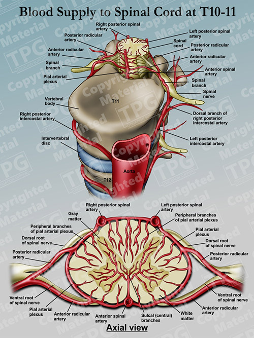
Blood Supply To Spinal Cord At T10-11 - Order
 presentationgroup.com
spinal cord blood supply t10 spine infection order staph sepsis result death innervation
Normal Anatomy Of The Ankle | Doctor Stock
 doctorstock.photoshelter.com
ankle anatomy anterior foot malleolus medial ligaments left lateral ligament leg bones lower joint talus labeled diagram anatomical syndesmosis human
Spinal Anatomy Chart - Clinical Charts And Supplies
 clinicalcharts.com
anatomy spine spinal chart poster human chiropractic posters charts dog bones vertebrae skeleton acupunctureproducts medical cervical column drawing anatomical effects
Subluxation – Premier Chiropractic Centre
 premierchiropracticcentre.com
spinal subluxation chart spine vertebral bones complex organs column nerves describes happens affected
Spinal-nerves - Saratoga Spine
 www.saratogaspine.com
spinal nerves spine anatomy saratoga
Temporal Bone
 www.fpnotebook.com
anatomy temporal bone
Instant anatomy. Normal anatomy of the ankle. Vertebrae typical vertebra vertebral kenhub ligaments joints